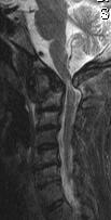
MRI scan of the spine
An MRI or Magnetic Resonance Imaging Scan can give a very clear picture of the structure of the spine, there by allowing your surgeon to look at the nerves, bones and soft tissues of your spine. The scan is extremely sensitive, but is only useful when the findings are interpreted in conjunction with a full history of the spinal condition and a thorough examination.
What is an MRI scan and is it safe?
The MRI Scan uses magnetic and radio waves, meaning that there is no exposure to X-rays or any other damaging forms of radiation. Radio waves stronger than the magnetic field of the earth are then sent through the body. This affects the body’s atoms, forcing the electrons into a different position. As they move back into place they send out radio waves of their own. The scanner picks up these signals and a computer turns them into a picture.
There are no known dangers or side effects connected to an MRI scan, however there is a small theoretical risk to the foetus during the first 12 weeks of pregnancy and ideally an MRI scans should be avoided during this period.
Do I need a scan?
The vast majority of patients with back pain can be treated following a careful history and examination of the spine. An MRI scan is not usually required, as this will not change the treatment for their back pain. A scan is however useful when there are symptoms of persistent nerve root irritation, causing pain in the arm or the leg, or for patients with severe persistent back pain in which injection therapy or surgery is being considered. On rare occasions back pain is caused by a serious condition such as a tumour, fracture or infection of the spine. Usually these conditions can be excluded with a thorough history and examination, however if a patient has “Red Flag Signs” then an MRI scan is required to exclude a serious condition.
What to expect if you are sent for a scan
 You will be asked to complete a questionnaire concerning the safe use of a scan. Some patients may not be suitable for a scan, reasons for this include people who are fitted with a cardiac pacemaker or have metal fragments in their eyes. These patients may then require a CT (Computerised Tomograpy) scan of the spine instead. Patients lie inside a large cylinder-shaped magnet, usually for about twenty minutes, while the scans are being made. The machine also makes a banging noise while it is working, which might be unpleasant, but usually music or ear defenders are available to limit this affect. Some people get claustrophobic during the test. Patients who are afraid this might happen should talk to the doctor beforehand, who may give them some medication to help them relax, or arrange for the MRI scan to be performed in a larger open scanner, although the images may be of a slightly poorer quality. Generally in the modern scanners people are able to enter feet first and the scanners are much shorter, both factors greatly reduce the feelings of claustrophobia. If you can keep very still the scan picture quality is better.
How will the scan help me?
 The scan does not tell us where back pain comes from, but may help your spinal specialist in localising the source of the pain and allowing treatment to be directed to the appropriate area of the spine. Prolapsed discs and bony fragments causing pressure on the nerves are easily seen on a MRI scan.


